Methods to microscopically observe
and identify asexual fungal structures
Purpose
The objectives of this laboratory exercise on September 12, 2012, were to 1) practice identifying species of Aspergillus by microscopically observing characteristic asexual fungal structures using tape and squash mounts learned during a previous laboratory exercise 2) learn to construct classical and modified Riddell mounts to microscopically observe intact asexual fungal structures 3) learn about different solid media to culture fungi for the unknowns project.
Materials and Methods
Fungal cultures of Aspergillus sp.
Microscope slides
Cover slips
Scalpel
Tweezers
Bent glass rods
Petri dishes
1/2 Potato Dextrose Agar medium
Dropper bottle with water
Spray bottle with 70% ethanol
Permanent marker
Paper towels
Metal striker
Bunsen burner
Canon PowerShot SD550 digital camera
Olympus CX31 compound microscope
A. Microscopic observation of Aspergillus sp.
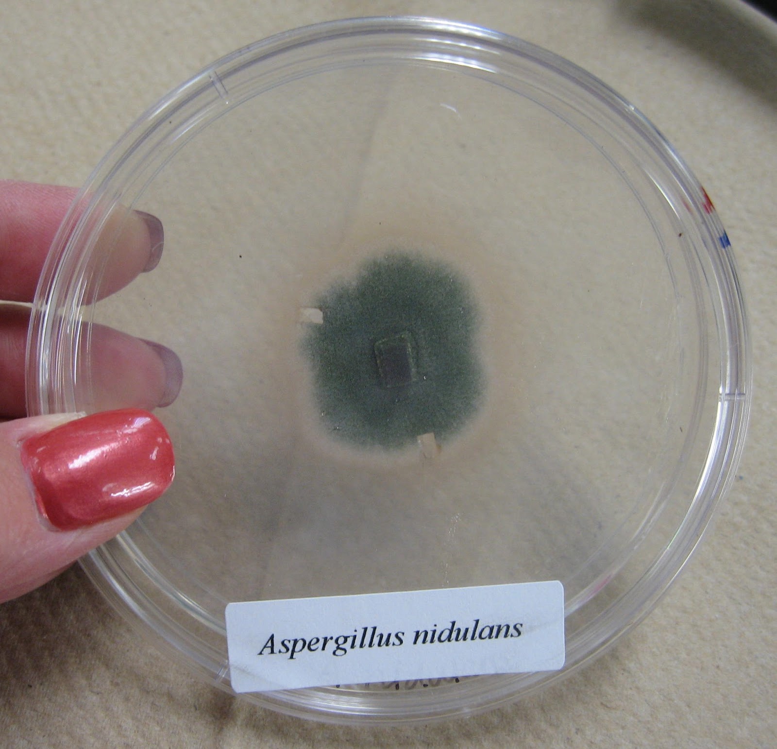
Tape and squash mounts learned during Lab 2 on September 5, 2012, were used to microscopically observe asexual fungal structures (uniseriate and biseriate vesicles, metulae, phialides, conidia, conidiophores) of A. niger, A. flavus, A. nidulans, A. oryzae, A. parasiticus, A. sojae and A. tamari. A Canon PowerShot SD550 digital camera was used to photograph fungal cultures and asexual structures through the eyepiece of an Olympus CX31 compound microscope that had been set to Köhler according to instructions also received during Lab 2.
B. Construction of classical and modified Riddell mounts
 Figure 1. Drawing detailing assembly of a classical Riddell mount to produce intact conidiophores for microscopic observation.
Figure 2. Drawing detailing assembly of a modified Riddell mount to produce intact conidiophores for microscopic observation.
To microscopically observe intact conidiophores, gently lift the cover slip off of the agar plug and place it fungal side down onto a drop of water on a microscope slide.
Figure 1. Drawing detailing assembly of a classical Riddell mount to produce intact conidiophores for microscopic observation.
Figure 2. Drawing detailing assembly of a modified Riddell mount to produce intact conidiophores for microscopic observation.
To microscopically observe intact conidiophores, gently lift the cover slip off of the agar plug and place it fungal side down onto a drop of water on a microscope slide.
C. Solid media to culture fungi
Dr. Brian Shaw discussed the composition and utility of various solid media available to the lab class to culture fungi for our unknowns project, including 1/2 Potato Dextrose Agar (PDA), water agar (WA), cornmeal agar, oatmeal agar, V8 agar and rose Bengal agar. 1/2 PDA enables faster conidiation. WA has fewer nutrients than 1/2 PDA, which allows for less dense growth and enables better microscopic observation.
Results
A. Microscopic observation of Aspergillus sp.
A. niger
 |
| Top of culture |
 |
| Bottom of culture |
 |
| Globose, dark brown, biseriate conidial head atop a smooth-walled, hyaline conidiophore (40X). Conidia are globose to subglobose, dark brown to black and rough walled. Photograph enlarged to show detail. |
 |
| Globose, dark brown, biseriate conidial heads atop smooth-walled, hyaline conidiophores (40X). Photograph cropped and enlarged to show detail. |
 |
| Classical Riddell mount showing colony formation of A. niger seven days after assembly. |
 |
| Vesicles forming atop conidiophores on cover slip (40X). Photograph cropped and enlarged to show detail. |
 |
| Mature conidial heads atop conidiophores protruding from edge of colony on cover slip (40X). Photograph cropped and enlarged to show detail. |
A. flavus
 |
| Classical Riddell mount showing colony formation of A. flavus seven days after assembly. |
 |
| Vesicles and conidiophores forming on cover slip (40X). Photograph cropped and enlarged to show detail. |
 |
| Vesicles and conidiophores forming on cover slip (40X). Photograph cropped and enlarged to show detail. |
 |
| Conidial heads, with chains of globose conidia, atop conidiophores protruding from edge of colony on cover slip (40X). Photograph cropped and enlarged to show detail. |
 |
| Conidial heads, with chains of globose conidia, atop conidiophores protruding from edge of colony on cover slip (40X). Photograph cropped and enlarged to show detail. |
 |
| Conidial heads, with globose conidia, atop of conidiophores on cover slip (40X). Photograph cropped and enlarged to show detail. |
A. parasiticus
.jpg) |
| Modified Riddell mount showing colony formation of A. parasiticus seven days after assembly. |
 |
| Conidial head, with chains of roughened conidia, atop a conidiophore on cover slip (40X). Photograph cropped and enlarged to show detail. |
 |
| Globose conidial head atop a conidiophore on cover slip (40X). Photograph cropped and enlarged to show detail. |
 |
| Conidiophore with vesicle on cover slip (40X). Photograph cropped and enlarged to show detail. |
 |
| Biseriate conidial heads atop conidiophores on cover slip (40X). Photograph cropped and enlarged to show detail. |
 |
| Conidial head atop a conidiophore on cover slip (40X). Photograph cropped and enlarged to show detail. |
 |
| Conidial head with chains of conidia atop a conidiophore on cover slip (40X). Photograph cropped and enlarged to show detail. |
Discussion
The arrangement of conidia on a conidiophore must be observed to properly identify a fungus to species. Although asexual fungal structures may be observed microscopically using tape and squash mounts, the Riddell mount is best for microscopically observing intact conidiophores. Both the classical and modified Riddell mounts produced intact conidiophores. However, I preferred the classical mount due to the fact that fungal structures were confined to cover slips, unlike in the modified mount. Morphological characteristics that are diagnostic for determining species of Aspergillus include uniseriate and biseriate vesicles, conidiophores and conidia (Figure 3).
Figure 3. Drawings of intact fungal conidiophores, showing morphological characteristics that are diagnostic for determining species of Aspergillus.
 Figure 1. Drawing detailing assembly of a classical Riddell mount to produce intact conidiophores for microscopic observation.


+culture.jpg)
+culture+%232.jpg)
.jpg)
.jpg)
+%232.jpg)


.jpg)
.jpg)
.jpg)


No comments:
Post a Comment